Fiocruz and Inmetro record images of coronavirus in action
07/10/2020
Maíra Menezes (IOC/Fiocruz)
In a partnership with the National Institute of Metrology, Quality, and Technology (Inmetro), researchers of the Oswaldo Cruz Institute (IOC/Fiocruz) have recorded new images that reveal the effects of the infection by the new coronavirus in the cells. Obtained through high-resolution microscopy, the photographs show that infected cells show membrane extensions, called filopodia, that form intercellular connections. This alteration may be one of the strategies of the virus to amplify infection.
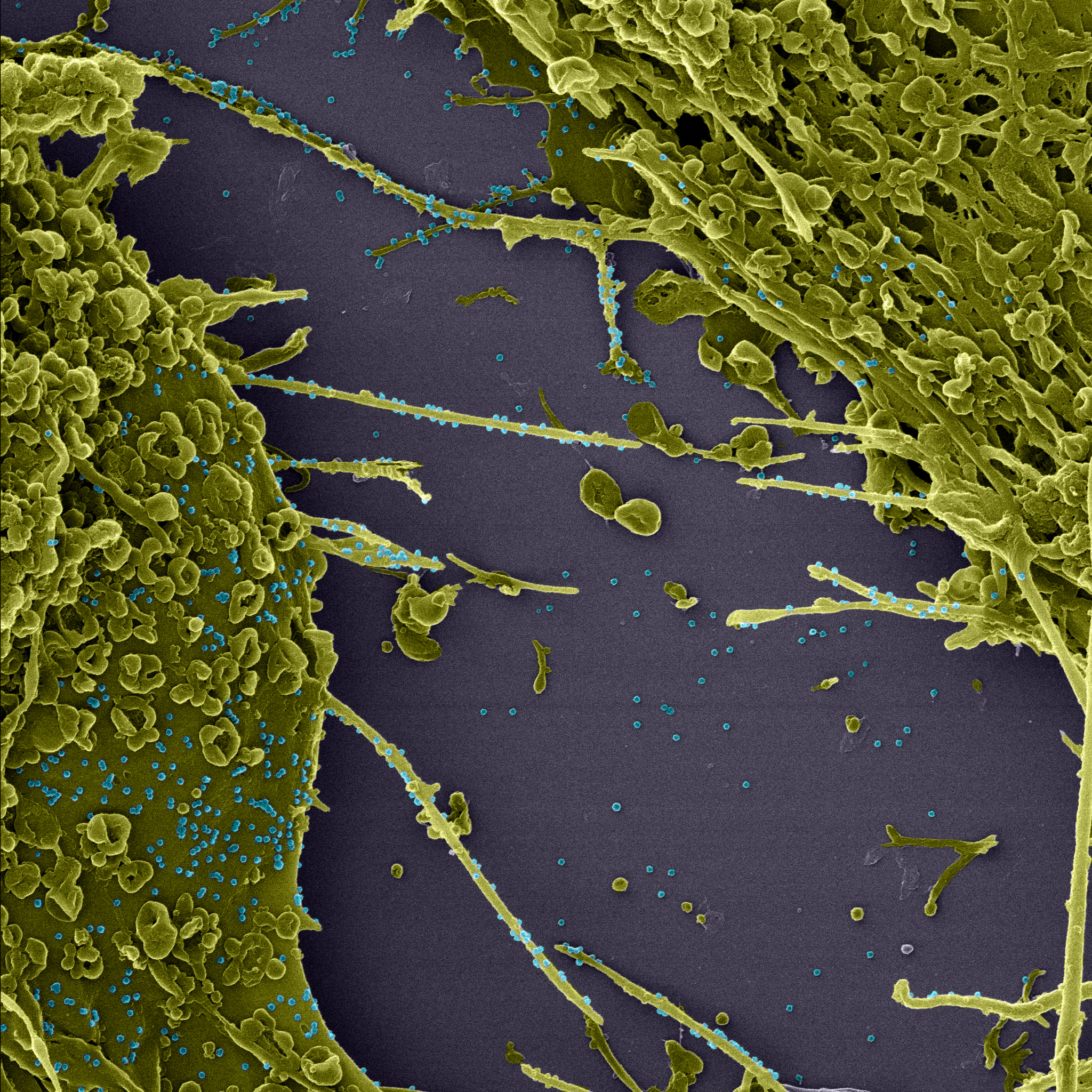
After 48 hours of infection with the new coronavirus, Vero cells have membrane extensions, which promote communication with neighboring cells. In blue, virus particles are observed on the cell surface (photo: LMMV / IOC / Fiocruz, LVRS / IOC / Fiocruz, and Nulam / Inmetro). Click on the image to enlarge
“Membrane extensions are part of the viral particles release process that replicates within the infected cells. They also promote intercellular communication, which we believe is related to the transference of viral particles to adjacent cells, thus maximizing the infection process”, explains researcher Debora Ferreira Barreto Vieira, of the Viral Morphology and Morphogenesis Laboratory of IOC/Fiocruz, coordinator of the research.
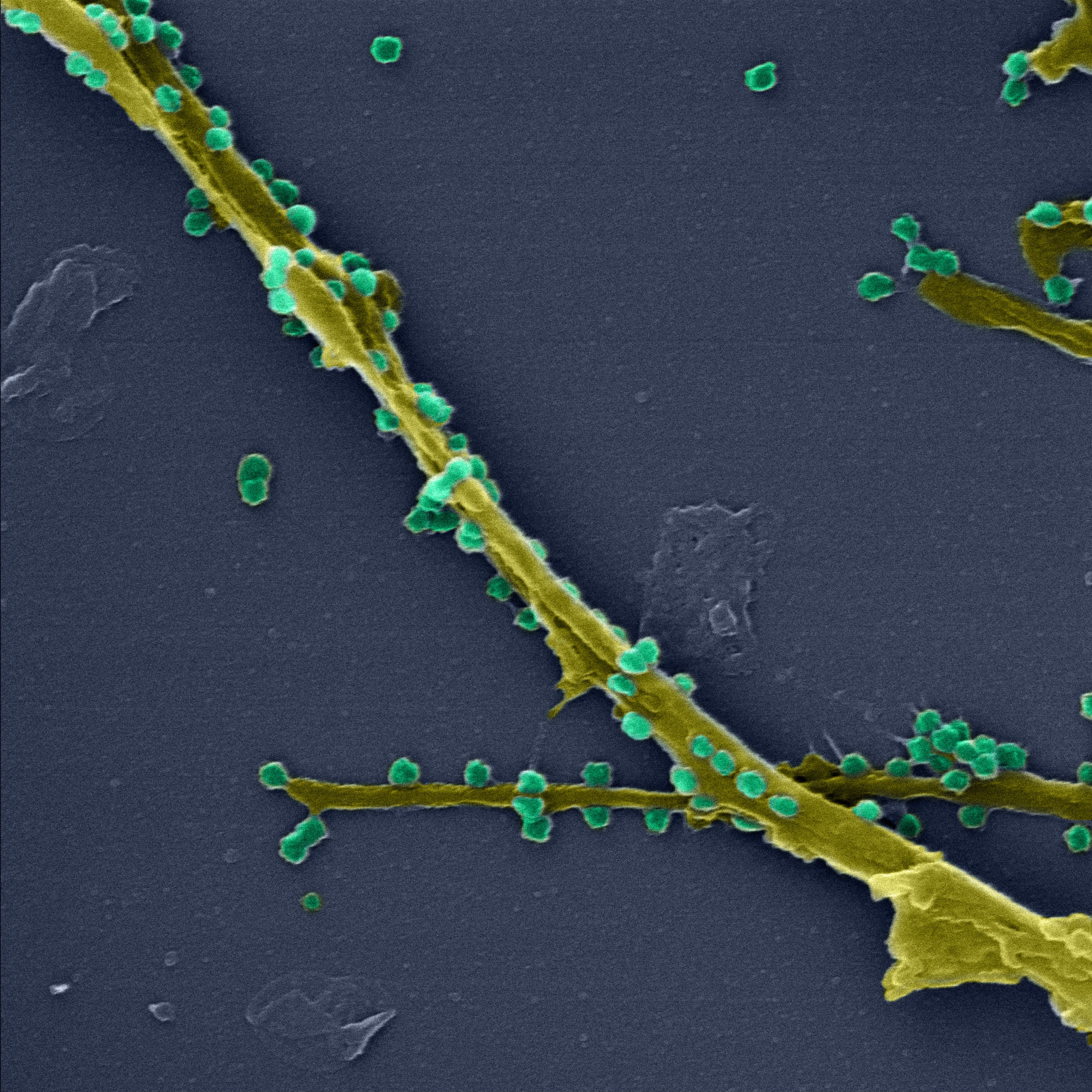
Microscopy imaging allows you to closely observe the connection between cells through membrane extensions and the presence of viral particles on the surface of the filopodia (photo: LMMV / IOC / Fiocruz, LVRS / IOC / Fiocruz, and Nulam / Inmetro). Click on the image to enlarge
The images, magnified up to 200 thousand times, show several membrane extensions in the infected cells, and the connection with neighboring cells. Many viral particles can be seen on the surface of the cell, including on the filopodia. According to Débora, these membrane extensions are caused by alterations in the cellular cytoskeleton caused by the infection. “There are very few viruses that cause this type of alteration in cells, such as ebola and Marburg. The process that makes these cells show this kind of alteration in infection by SARS-CoV-2 is an issue that is still open and we want to investigate”, says the researcher.
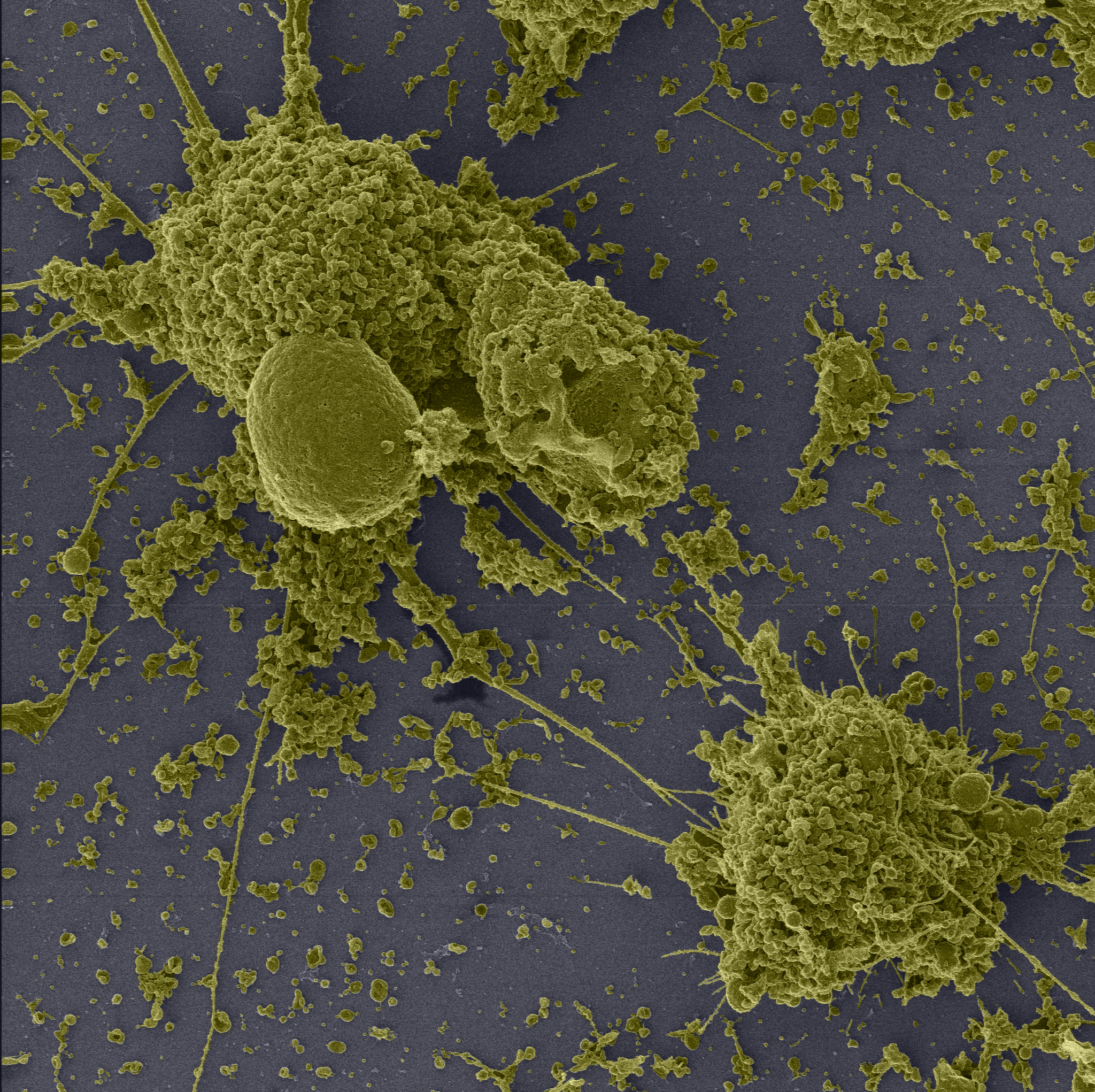
The photograph shows the marked change in cell structure after 72 hours of infection by the new coronavirus, including the presence of numerous filopodia (photo: LMMV / IOC / Fiocruz, LVRS / IOC / Fiocruz, and Nulam / Inmetro). Click on the image to enlarge
The study was carried out on Vero cells derived from monkey kidneys and very commonly used in virology studies. The images are the preliminary results of the research project “Study of the morphology, morphogenesis, and pathogenesis of the Sars-CoV-2 in systems in vitro, in vivo”, made in cooperation with the Laboratory of Viral Morphology and Morphogenesis and the Laboratory of Respiratory Viruses and Measles of the IOC. The photographs were recorded with a triple-beam high-resolution microscope, in cooperation with researchers Braulio Archanjo and Willian Silva, of Inmetro’s Laboratory of Electronic Microscopy.