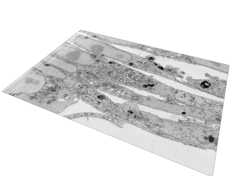
Animation presents a three-dimensional reconstruction of the 'viral factory' in a cell infected by the mpox virus: dark blue represents the cell nucleus; orange represents mature viral particles; green represents mitochondria; yellow represents rough endoplasmic reticulum; violet represents empty vesicles; purple represents immature viral particles; and light blue represents viral DNA (Image: Barreto-Vieira and collaborators [1])
Researchers from the Oswaldo Cruz Institute (IOC/Fiocruz) have just presented another important contribution to global science: pioneering advances in understanding the process of infection and replication of the mpox virus (MPXV), which has been expanding its global presence since 2022.
Formerly known as monkeypox, and renamed by the World Health Organization (WHO) to overcome the stigma associated with the disease, mpox is characterized by skin lesions (such as red spots, small sores, or blisters), usually concentrated on the face, palms of the hands, and soles of the feet. These lesions can also appear in the mouth, eyes, genitals, and anus.
The WHO recently recognized two international public health emergencies associated with the pathogen: the first between July 2022 and May 2023 and the second from August 2024.
One of the countries most affected by the disease, Brazil has recorded more than 13,000 cases since 2022, with near 2,000 cases in 2024 and around 100 in 2025. Sixteen deaths have been confirmed in the country.
Using microscopy records and three-dimensional modeling, the study published in the Journal of Medical Virology details the step-by-step process of the viral infection in mammalian cells, contributing to an understanding of what happens in the human body.
The study obtained the first images of the process of mpox entering cells. It also presents a groundbreaking 3D animation model that shows the structures of the 'viral factory' that forms inside the cell for the virus replication. Further, it reveals images of the release of viral particles.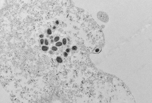
Transmission electron microscopy image shows viral particles at the cell periphery. One particle appears to be in the process of extrusion, inside an exocytosis vesicle (image: Barreto-Vieira and collaborators)
Debora Ferreira Barreto-Vieira, head of the IOC/Fiocruz's Laboratory of Viral Morphology and Morphogenesis, emphasizes that the study fills gaps in the scientific literature.
"Mpox was isolated from monkeys in the 1950s and from humans in the 1970s. Despite all this time, when the first public health emergency was declared in 2022, there was little published data on the replicative cycle of the virus. Our findings expand knowledge in the field of virology and could help in the development of antiviral therapies and infection prevention measures," says Débora.
The work was carried out by IOC's Viral Morphology and Morphogenesis Laboratory in partnership with the Microscopy Laboratories Center of the National Institute of Metrology, Quality and Technology (Nulam/Inmetro), led by researcher Bráulio Soares Archanjo.
Step-by-step infection
For the research, the scientists infected Vero cells, which come from monkey kidneys and are widely used in virology studies, with a strain of the mpox virus isolated from a sample of a patient in Rio de Janeiro in 2022.
The sample was provided by IOC's Laboratory of Respiratory Viruses, Exanthematics, Enteroviruses and Viral Emergencies, which acts as a diagnostic reference for the Ministry of Health.
The study identified that mpox enters cells through the process of endocytosis, in which the cell membrane surrounds the viral particle, forming a vesicle, which is then internalized.
The virus then reaches the cytoplasm, where it mobilizes cellular structures for replication, creating a "viral factory" near the nucleus of the cell.
From approximately 1,200 images captured using the focused ion beam (FIB) scanning electron microscopy technique, the scientists produced a three-dimensional model of this structure.
The work showed that the virus replicates in an area of the cell surrounded by mitochondria (structures responsible for energy production) and cisternae of the rough endoplasmic reticulum (an organelle that synthesizes and modifies proteins). In this area, numerous empty vesicles and viral particles in different stages of formation can be seen.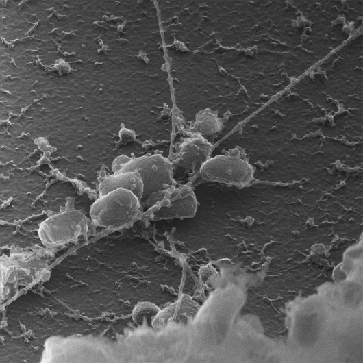
Scanning electron microscopy image shows viral particles attached to a filament of the cytoskeleton (image: Barreto-Vieira and collaborators)
Another relevant finding was the recording of images showing the adhesion of viral particles to cytoskeletal filaments, which are protein fibers responsible for the structure of cells. These filaments drive the transport of newly formed viruses inside the cell, facilitating their release into the outside environment.
The process had already been identified by transmission electron microscopy in previous studies and was documented in detail through scanning electron microscopy.
One of the records of this process, with artistic color, ranked 2nd in the Photography Award - Science and Art, promoted by the National Council for Scientific and Technological Development (CNPq).
The research also revealed that the mpox leaves the cell through the process of exocytosis, in which vesicles containing viral particles fuse with the cell membrane, causing the viruses to be released while the cell remains intact.
The infection causes various types of damage to the cells, leading to cell death within a few days, as documented in the study and in a previous research by the Institute.
Mpox
The mpox virus belongs to the Orthopoxvirus genus, of the Poxviridae family, which also includes the pathogens responsible for human and bovine smallpox, as well as the vaccinia virus, used in the production of the vaccine that eradicated human smallpox.
The pathogen became an international public health emergency for the second time in August last year.
The emergency scenario, currently ongoing, was declared by the WHO due to the outbreak in the Democratic Republic of Congo and neighboring countries caused by a potentially more aggressive strain of mpox, called clade 1b.
The first declaration of emergency associated with the virus occurred in July 2022, when another viral strain, known as clade 2, spread to over a hundred countries. This emergency ended in May 2023, considering the reduction in cases of the disease, although the virus continues to circulate at low levels.
According to the WHO, from January 2022 to December 2024, 124,000 cases and 272 deaths from the disease were confirmed in 128 countries.
In addition to skin lesions, the main symptoms of the disease are swollen lymph nodes (lymphadenopathy), fever, weakness, headaches, and body aches.
The Ministry of Health advises people with suspected symptoms of the infection to seek a health unit for evaluation. The treatment aims to alleviate symptoms, as there is still no available antiviral medication.
Although most cases of mpox are mild or moderate, some individuals have a higher risk of developing severe forms. Vaccination is indicated as prevention for people living with HIV and professionals who work directly with Orthopoxvirus in laboratories.
Immunization may also be indicated for people who have had contact with secretions or fluids from patients with a suspected or confirmed diagnosis of mpox, as evaluated by the local surveillance team.